By: Katelyn Thickett
This is a blog post aimed at the general public that is broken down and described at a basic level of knowledge of cognitive impairments and imaging techniques used. My goal is to describe why neurological testing is used in regard to cognitive impairments, what types of testing are used, and when these tests are used.
To begin, let me break down what neurological testing is and what cognitive impairments include. To simplify, cognitive is anything to do with the brain and what is happening within it. Thus, cognitive techniques include different imaging that looks at the brain and different structures as well as activity within the brain. Cognitive impairment is anything that alters the normal functioning of the brain concerning learning, knowing, and understanding.
Cognitive impairments arising from the following medical issues will be the focus in this article:
- Alzheimer’s and Dementia
- Attentional Deficit Hyperactivity Disorder (ADHD)
- Autism Spectrum Disorder (ASD)
- Brain Tumors (benign and malignant)
- Multiple Sclerosis (MS)
- Parkinson’s Disease
- Seizures (Epileptic and non-epileptic)
- Sleep Disorders
- Stroke (sometimes can cause cognitive impairment)
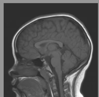
This MRI image is of Katelyn Thickett’s brain in 2010.
Dementia is an umbrella term for brain disease whereas Alzheimer’s is a specific type of dementia. These result in interference with memory as well as other functioning sometimes involving problems with language and problem-solving.
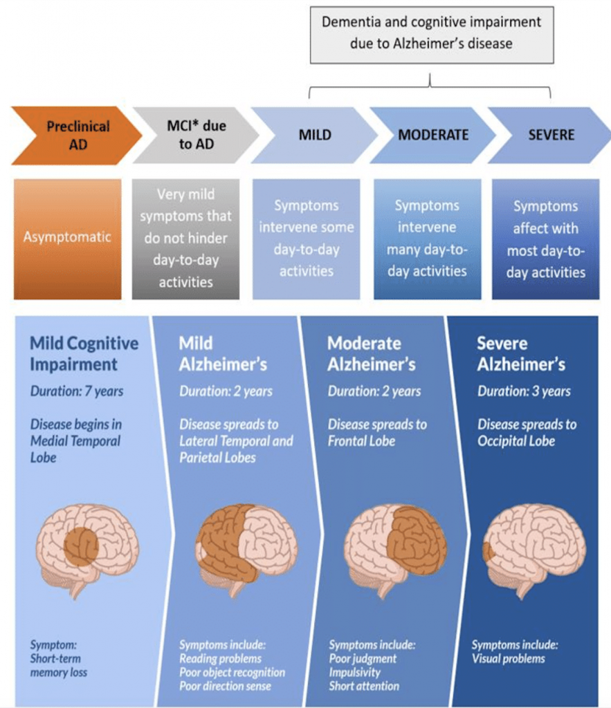
(Dan, 2021)
Attentional Deficit Hyperactivity Disorder (ADHD) is a neurodevelopmental disorder, characterized by hyperactivity, inattention, and impulsivity that can interfere with functioning and development.
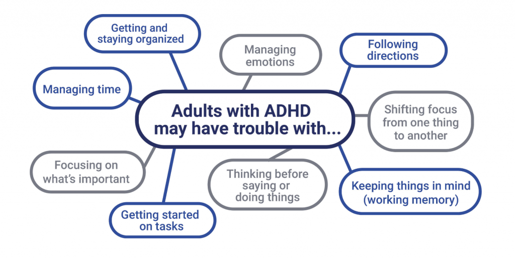
(Gharibian, n.d.)
Autism Spectrum Disorder (ASD) is a developmental disorder that can affect different aspects of the brain that oversee social interaction, communication, and learning.
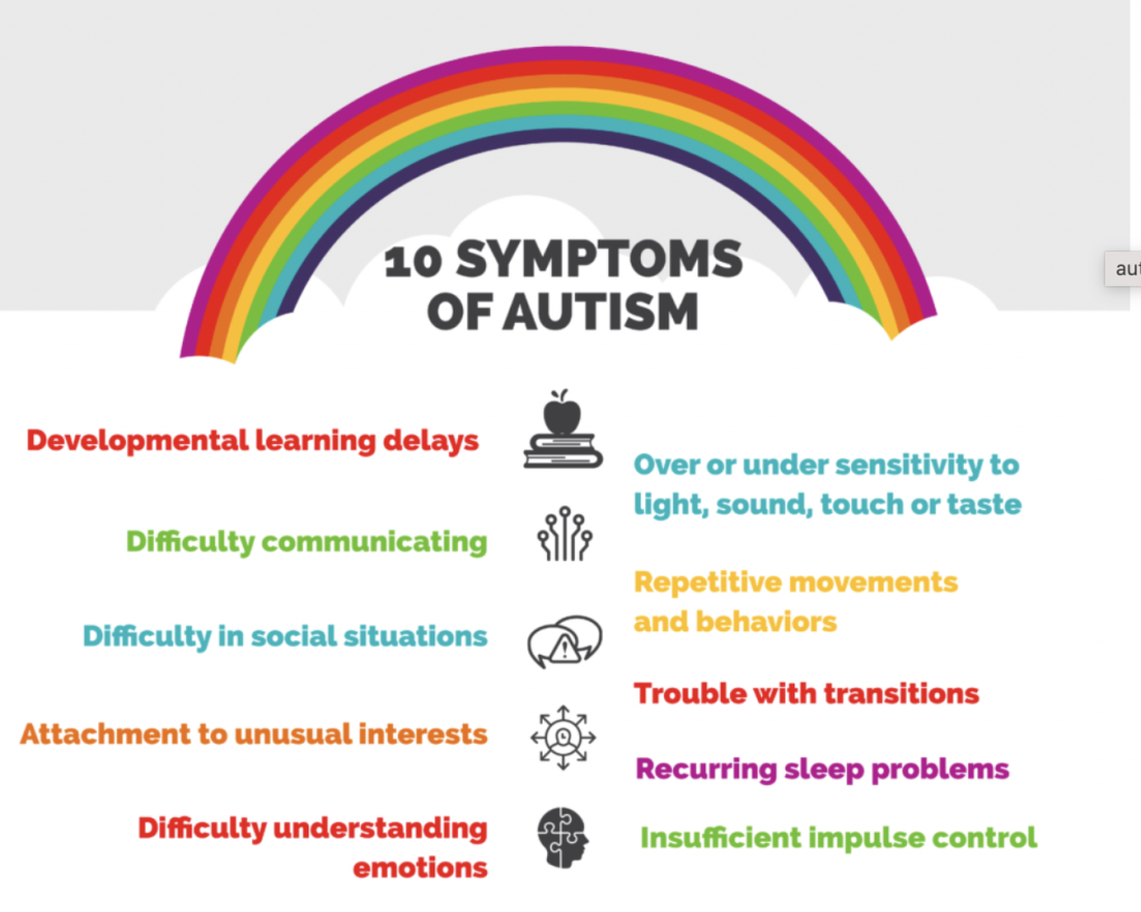
(Applied Behaviour Analysis Programs Guide, 2022)
Brain Tumors (cancerous and non-cancerous) are clusters of abnormal cells within the brain, malignant meaning cancerous and benign meaning non-cancerous. The location of a tumor determines the effects it may have on different cognitive aspects. Below is a photo that contains the parts and the effects of different brain tumors.
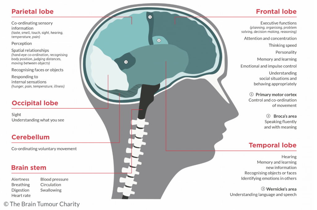
(The Brain Tumour Charity, n.d.)
Multiple Sclerosis (MS) is the breaking down of the myelin which is the fatty tissue that insulates the neural connections within the brain. The deterioration of the fatty tissue results in the slowing down of brain activity or in some cases the inability for brain activity. In simple terms, MS is an autoimmune disease that is characterized by brain lesions (abnormal brain tissue).
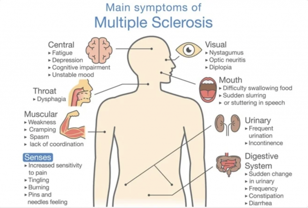
(Cheriyedath, 2021)
Parkinson’s Disease is an ongoing brain disease that affects bodily movements due to the destruction of dopamine which is a neurotransmitter (messengers within the brain).
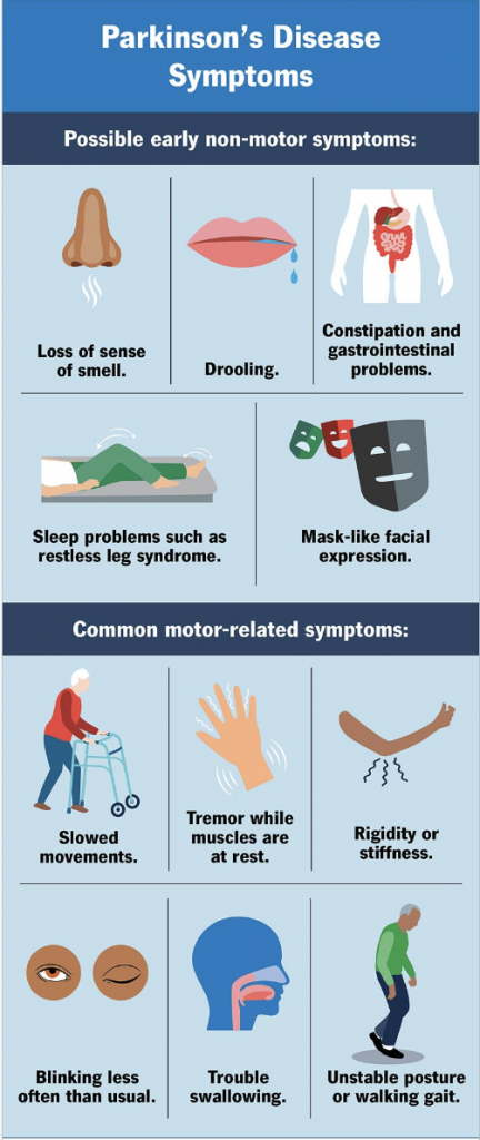
(Cleveland Clinic, 2022)
Seizures (Epileptic and non-epileptic) Epileptic seizures indicate abnormal bursts of brain activity whereas non-epileptic seizures do not contain abnormal brain activity. Both can affect different functions including movement and speech. Below are the types of epileptic seizures and how they present.
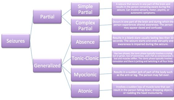
(Google, n.d.)
Sleep Disorders are abnormal patterns during sleep that affect normal brain functioning.

(Sunshine Behavioral Health, 2024)
Stroke (sometimes can cause cognitive impairment) indicates a blood clot (ischemic stroke) or a rupture of blood vessels (hemorrhagic stroke) within the brain. While the pre-stroke symptoms tend to be consistent, the post-stroke outcomes vary significantly depending on the location of the stroke within the brain.
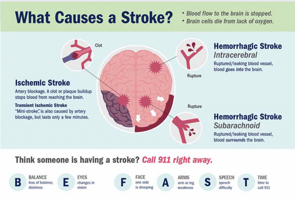
(Rivera, 2022)
Neurological Testing that will be discussed in this article will include:
- Computerized Tomography (CT scan)
- Diffusion Tensor Imaging Tractography (DTI)
- Electroencephalogram (EEG)
- Functional Magnetic Resonance Imaging (fMRI)
- Magnetic Resonance Imaging (MRI)
- Positron Emission Tomography (PET)
Computerized Tomography (CT) is also known as a “CAT scan” and is like a big three-dimensional X-ray machine that can take photos of the entire body including the brain (Reisburg, 2021). CT scans tend to be more readily available as well as cheaper and quicker than other imaging techniques like an MRI. Although CT scans may not be as detailed as an MRI, they tend to be the first referral for cognitive impairments. CT scans are not recommended to get often as they do let off radioactivity which in great amounts can cause health concerns.
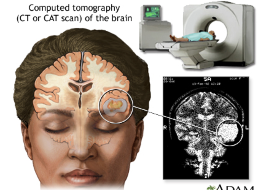
(Medline Plus, 2023)
- Generally, a CT scan is the first imaging used when a brain tumor is suspected, as it provides a basic structural image of the brain in a timely manner as well as potential surgical planning of the location (Bruzzone et al., 2012)
- Sometimes CT scans are used after a seizure to determine if there are any obvious structural changes within the brain (Kuzniecky, 2013).
- A quick way to identify the type of stroke and the location within the brain. Treatment of stroke promptly is crucial for the best outcome (Birenenum et al., 2011)
- A CT scan could be used to rule out other possible causes of symptoms related to multiple sclerosis, the CT scan itself is not detailed enough to indicate lesions caused by MS but could be used to rule out other possible causes (Pietrangelo, 2018).
Diffusion Tensor Imaging Tractography (DTI) is an MRI technique that indicates the diffusion of water and its direction within the brain (Reisburg, 2021).
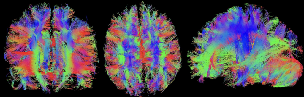
(Woodard, 2020)
- Used in ADHD, DTI indicates the lack of neuro-networks responsible for attention and impulse control (Bouziane et al., 2017).
- For MS, DTI displays the little changes that are within the white matter (brain tissue abnormality), this is correlated with cognitive dysfunction severity (Sbardella et al., 2013).
- A DTI displays the damage and changes of white matter following a stroke (Pinter et al., 2020).
Electroencephalogram (EEG) is a device that records brain activity using electrodes placed on the scalp (Reisburg, 2021).
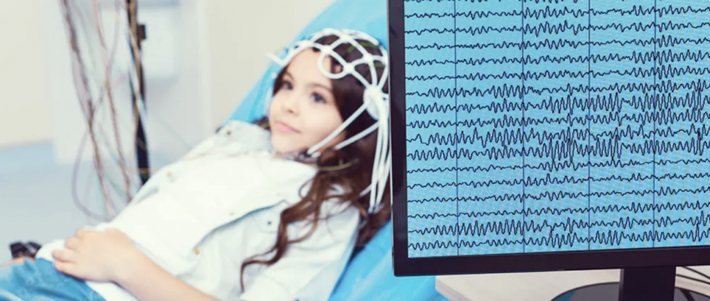
(Alivio Medical Center, 2022)
- Used in locating abnormal brain activity in individuals with seizures. Some types of seizures do not involve abnormal brain activity (non-epileptic) and therefore can be distinguished using an EEG (Ein et al., 2023).
- Used to detect and monitor sleep disorders in recording brain activity during different stages of sleep (Behzad & Behzad, 2021).
Functional Magnetic Resonance Imaging (fMRI) is a three-dimensional imagining technique that uses magnetic fields to develop images that display the small changes in blood flow during brain activity (Reisburg, 2021).
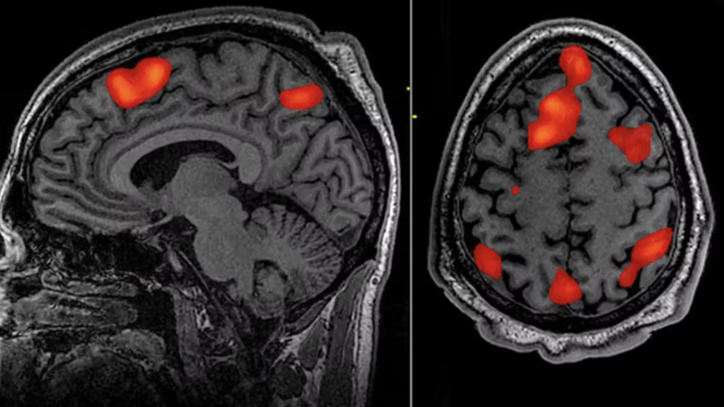
(PsychCentral, 2021)
- An fMRI can show abnormal activation patterns within areas of the brain responsible for attention and executive function related to ADHD (Paloyelis et al., 2007). Executive control includes the mental resources that are responsible for setting goals, setting priorities as well as avoiding conflict (Reisburg, 2021).
- In Autism Spectrum Disorder, an fMRI has shown abnormal activation patterns during social tasks (interpersonal skills) (Kim et al., 2015).
- For Parkinson’s disease, fMRI has displayed abnormal activation within the brain during cognitive tasks (Filippi et al., 2019). Some cognitive tasks that are commonly included with Parkinson’s Disease may include bradykinesia (slowed movements), rigidity (stiff movements), and/or tremor at rest (Filippi et al., 2019).
- An fMRI can be used post-stroke to map the functional reorganization of the brain (Golestani et al., 2013). For example, if the left hemisphere of the brain is disrupted in the stroke, language and communication abilities may be disrupted resulting in aphasia (disruption in language capabilities) (Reisburg, 2021). If Broca’s area (responsible for the production of speech)is disrupted, non-fluent aphasia (the inability to speak or write with fluency) can occur (Reisburg, 2021).
- The use of a resting state fMRI can display disruptions within connections of brain activity that are responsible for cognitive decline in Multiple Sclerosis (Benedict et al., 2020). Cognitive processing speed (how long it takes for someone to do something) and episodic memory (long-term memory system that is in charge of remembering specific events) are a few regions where decline is seen within MS (Benedict et al., 2020).
- An interesting study discusses the use of an MRI in distinguishing between the thicknesses of various brain regions in those with mild cognitive impairment versus those with Alzheimer’s disease (Desikan et al., 2009). The results indicate that an automated MRI program can display the brain differences between those with Alzheimer’s and those with normal cognitive decline that occurs naturally with aging (Desikan et al., 2009). This could be potentially useful in acting as an additional resource to see to what extent biologically an individual’s cognitive impairments are currently.
Magnetic Resonance Imaging (MRI) is a three-dimensional imagining technique that uses magnetic fields to develop images that display brain structures and their function (Reisburg, 2021).
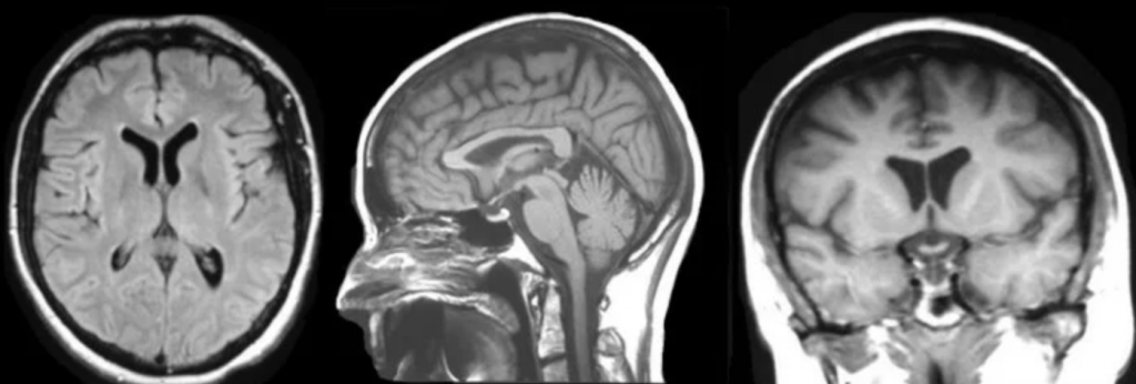
(Preston, 2006)
- In Dementia, an MRI can show brain abnormalities including brain atrophy (loss of brain cells and connections) (Desikan et al., 2009).
- For brain tumors, an MRI provides a detailed image as well precise location of the tumor (Bruzzone et al., 2012).
- In Multiple Sclerosis, MRIs are used to see brain lesions (brain abnormalities) that are related to cognitive dysfunction severity (Benedict et al., 2020)
- In Parkinson’s Disease, an MRI displays structural changes that are related to the movement and emotional areas of the brain that are affected in PD (Srakocic, 2022).
Positron Emission Tomography (PET) is the measure of glucose (the brain’s fuel) within brain regions which indicates the energy within that brain region (Reisburg, 2021).
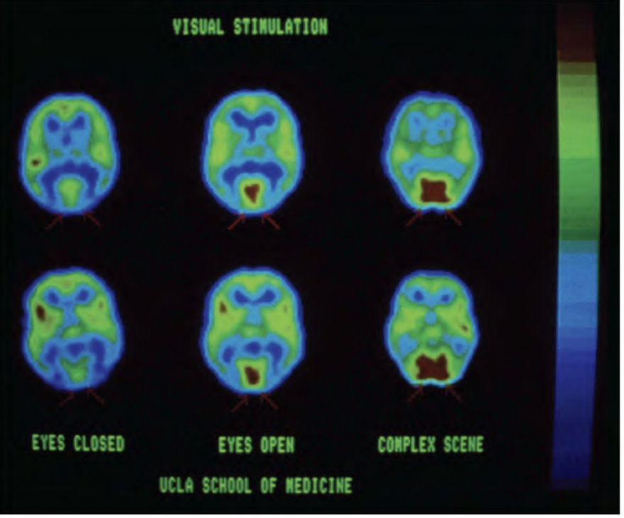
(Reisberg, 2021 p. 38)D
- Used specifically for cancerous brain tumors to give an estimate of the severity of the cancer (Chen, 2007).
- PET can display the plaque deposits on the brain that are associated with dementia and are linked to cognitive decline in dementia (Marcus et al., 2014). Cognitive decline within dementia may include problems with learning, memory, and language.
- For epileptic seizures, a PET can display the networks within the brain that can be the cause of the seizures (Cottage Health, 2024).
A Few Things to Keep in Mind…
- The brain is a complex structure that we do not fully understand yet despite neuroimaging techniques.
- Brain abnormalities can vary in severity and therefore symptoms can vary greatly even within the same diagnosis.
- The brain is always going through changes and is constantly changing.
- Brain impairments vary depending on the location of abnormality (different parts of the brain, operate different functions).
- Sometimes ongoing and a variety of neurological testing is needed to see the progress of brain abnormalities.
- Neurological testing is not a treatment but aids in diagnosing brain abnormalities.
- It is important to remember that the left hemisphere of the brain governs the movements and functions of the right side of the body, and vice versa.
About the Author: Katelyn Thickett is in her third year at Capilano University in North Vancouver, BC. Her interests include neuropsychology, clinical psychology, and music and the brain. Katelyn loves hiking and anything to do with the outdoors as well as travelling to new places. Some of Katelyn’s goals include pursuing a master’s in psychology and eventually a registered psychologist.
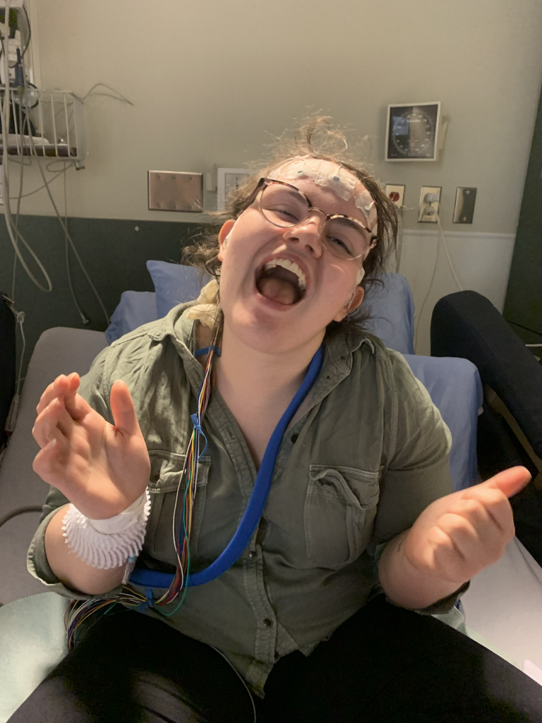
This photo is Katelyn Thickett in Foothills seizure monitoring unit hooked up to an EEG machine in 2019.
References
Alivio Medical Center. (2022). Electroencephalogram EEG [Image]. Retrieved from https://aliviohealth.com/what-is-an-electroencephalogram-eeg/
Applied Behavior Analysis Programs Guide. (2022). Ten symptoms of Autism [Diagram]. Retrieved from https://www.appliedbehavioranalysisprograms.com/lists/5-symptoms-of-autism/
Behzad, R. and Behzad, A. (2021) The Role of EEG in the Diagnosis and Management of Patients with Sleep Disorders. Journal of Behavioral and Brain Science, 11, 257-266. doi: 10.4236/jbbs.2021.1110021.
Benedict, R. H. B., Amato, M. P., DeLuca, J., & Geurts, J. J. G. (2020). Cognitive impairment in multiple sclerosis: clinical management, MRI, and therapeutic avenues. The Lancet. Neurology, 19(10), 860–871. https://doi.org/10.1016/S1474-4422(20)30277-5
Birenbaum, D., Bancroft, L. W., & Felsberg, G. J. (2011). Imaging in acute stroke. The western journal of emergency medicine, 12(1), 67–76.
Bouziane, C., Caan, M. W. A., Tamminga, H. G. H., Schrantee, A., Bottelier, M. A., de Ruiter, M. B., Kooij, S. J. J., & Reneman, L. (2017). ADHD and maturation of brain white matter: A DTI study in medication naive children and adults. NeuroImage. Clinical, 17, 53–59. https://doi.org/10.1016/j.nicl.2017.09.026
Bruzzone, M. G., D’Incerti, L., Farina, L. L., Cuccarini, V., & Finocchiaro, G. (2012). CT and MRI of brain tumors. The quarterly journal of nuclear medicine and molecular imaging : official publication of the Italian Association of Nuclear Medicine (AIMN) [and] the International Association of Radiopharmacology (IAR), [and] Section of the Society of…, 56(2), 112–137.
Chen, W. (2007). Clinical Applications of PET in Brain Tumors. Journal of Nuclear Medicine, 48(9), 1468-1481. https://doi.org/10.2967/jnumed.106.037689
Cheriyedath, S. (2021). Main symptoms of Multiple Sclerosis [Diagram]. Retrieved from https://www.news-medical.net/health/Early-signs-of-Multiple-Sclerosis.aspx
Cleveland Clinic. (2022). Parkinson’s Disease symptoms [Diagram]. Retrieved from https://my.clevelandclinic.org/health/diseases/8525-parkinsons-disease-an-overview
Cottage Health. (2024). EEG monitored PET scan – what to expect. Retrieved from https://www.cottagehealth.org/services/epilepsy/eeg-monitored-pet-scan/
Dan, S. (2021). The progression of Alzheimer’s disease from brain changes [Diagram]. Retrieved from https://www.researchgate.net/figure/The-progression-of-Alzheimers-disease-from-brain-changes_fig1_351118963
Desikan, R. S., Cabral, H. J., Hess, C. P., Dillon, W. P., Glastonbury, C. M., Weiner, M. W., Schmansky, N. J., Greve, D. N., Salat, D. H., Buckner, R. L., & Fischl, B. (2009). Automated MRI measures identify individuals with mild cognitive impairment and Alzheimer’s disease. Brain, 132(8), 2048–2057. https://doi.org/10.1093/brain/awp123
Ein Shoka, A. A., Dessouky, M. M., El-Sayed, A., & Hemdan, E. E. (2023). EEG seizure detection: concepts, techniques, challenges, and future trends. Multimedia tools and applications, 1–31. Advance online publication. https://doi.org/10.1007/s11042-023-15052-2
Filippi, M., Sarasso, E., & Agosta, F. (2019). Resting-state Functional MRI in Parkinsonian Syndromes. Movement disorders clinical practice, 6(2), 104–117. https://doi.org/10.1002/mdc3.12730
Gharibian, E. (n.d.). Adults with ADHD may have trouble with… [Diagram]. Retrieved from https://verdugopsych.com/what-is-adhd-an-overview-of-the-causes-and-signs-of-adhd/
Golestani, A. M., Tymchuk, S., Demchuk, A., Goodyear, B. G., & VISION-2 Study Group (2013). Longitudinal evaluation of resting-state FMRI after acute stroke with hemiparesis. Neurorehabilitation and neural repair, 27(2), 153–163. https://doi.org/10.1177/1545968312457827
Google (n.d.). Flowchart of seizure types [Diagram]. Retrieved from https://www.pinterest.ca/pin/551972498070258819/
Kim, S. Y., Choi, U. S., Park, S. Y., Oh, S. H., Yoon, H. W., Koh, Y. J., Im, W. Y., Park, J. I., Song, D. H., Cheon, K. A., & Lee, C. U. (2015). Abnormal activation of the social brain network in children with autism spectrum disorder: an FMRI study. Psychiatry investigation, 12(1), 37–45. https://doi.org/10.4306/pi.2015.12.1.37
Kuzniecky, R. (2013). Brain imaging for epilepsy. Epilepsy Foundation. https://www.epilepsy.com/diagnosis/brain-imaging
Marcus, C., Mena, E., & Subramaniam, R. M. (2014). Brain PET in the diagnosis of Alzheimer’s disease. Clinical nuclear medicine, 39(10), e413–e426. https://doi.org/10.1097/RLU.0000000000000547
Medline Plus. (2023). CT scan of the brain [Image]. Retrieved from https://medlineplus.gov/ency/imagepages/19237.htm
Paloyelis, Y., Mehta, M. A., Kuntsi, J., & Asherson, P. (2007). Functional MRI in ADHD: a systematic literature review. Expert review of neurotherapeutics, 7(10), 1337–1356. https://doi.org/10.1586/14737175.7.10.1337
Pietrangelo, A. (2018). Multiple sclerosis and radiology: What you should know. Healthline. https://www.healthline.com/health/multiple-sclerosis/multiple-sclerosis-radiology#radiology
Pinter, D., Gattringer, T., Fandler-Höfler, S., Kneihsl, M., Eppinger, S., Deutschmann, H., Pichler, A., Poltrum, B., Reishofer, G., Ropele, S., Schmidt, R., & Enzinger, C. (2020). Early Progressive Changes in White Matter Integrity Are Associated with Stroke Recovery. Translational stroke research, 11(6), 1264–1272. https://doi.org/10.1007/s12975-020-00797-x
Preston, D. (2006). Magnetic Resonance Imaging [Image]. Retrieved from https://case.edu/med/neurology/NR/MRI%20Basics.htm
PsychCentral. (2021). Functional Magnetic Resonance Imaging [Image]. Retrieved from https://psychcentral.com/lib/what-is-functional-magnetic-resonance-imaging-fmri
Reisberg, D. (2021). Cognition exploring the science of the mind (8th ed.). W.W. Norton & Company.
Rivera, L. S. (2022). What causes a stroke [Diagram]. Retrieved from https://blog.uvahealth.com/2022/10/06/how-different-types-strokes-happen/
Sbardella, E., Petsas, N., Tona, F., & Pantano, P. (2015). Resting-State fMRI in MS: General Concepts and Brief Overview of Its Application. BioMed research international, 2015, 212693. https://doi.org/10.1155/2015/21269
Sbardella, E., Tona, F., Petsas, N., & Pantano, P. (2013). DTI Measurements in Multiple Sclerosis: Evaluation of Brain Damage and Clinical Implications. Multiple sclerosis international, 2013, 671730. https://doi.org/10.1155/2013/671730
Srakocic, S. (2022). Parkinson’s MRI: Diagnosing early onset through brain imaging. Healthline. https://www.healthline.com/health/parkinsons/parkinsons-mri#takeaway
Sunshine Behavioral Health. (2024). Types of sleep disorders [Diagram]. Retrieved from https://sunshinebehavioralhealth.com/resources/sleep-mental-health/
The Brain Tumour Charity. (n.d.). The human brain [Diagram]. Retrieved from https://www.thebraintumourcharity.org/brain-tumour-signs-symptoms/brain-tumour-location-symptoms/
Woodard, B. (2020). Diffusion Tensor Imaging [Image]. Retrieved from https://www.porterrennie.com/blog/diffusion-tensor-imaging-traumatic-brain-injury
Leave a Reply